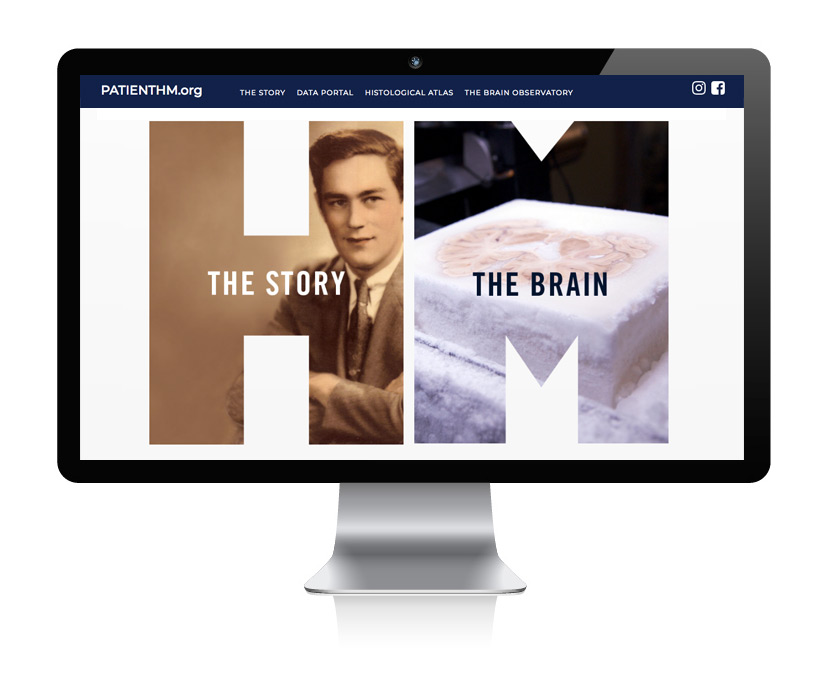
The human brain, up close and personal
We study neurological patients who changed the way we understand the human brain.
The scientific mission of The Brain Observatory is to build an interactive library of brain images and models that can support research, medical education, and drug development.
Our goal is to catalog the stories and neuroimaging data from as many human brains as possible so that we can reveal what makes us who we are. We use a fleet of motorized, digitizing microscopes to visualize and measure the human brain at cellular resolution. By linking microscopic features to each patient’s unique experience we can advance diagnostic precision and support personalized, preventive healthcare for the brain.
Some of the patients whose case was studied at The Brain Observatory have changed neuroscience history or are challenging current theories of brain function, like patients H.M. and A.V.
The life and brain of Henry G. Molaison
Researchers at The Brain Observatory examined the brain of Henry G. Molaison, the most important medical case in neuroscience history. The results and all data from the our study are freely available for researchers and educators at PatientHM.org.
Henry G. Molaison was a young man who, in the summer of 1953, became profoundly amnesic after an experimental surgery against his severe epileptic seizures. The surgery was directed at the Medial Temporal Lobes (MTL) of Henry’s brain with the goal of removing the hippocampus and neighboring structures. That is when Henry became Patient H.M. who would, for the rest of his life, take part in scientific experiments that fundamentally advanced our understanding of how memory works.
Henry G. Molaison (left) and Dr. Scoville (right)
Patient H.M. died December 2nd, 2008. Dr. Annese and his research team at The Brain Observatory were selected and awarded a grant from the National Science Foundation to examine his brain postmortem. Using a novel procedure developed at The Brain Observatory that combined physical dissection and 3D imaging researchers created a microscopic model of the whole brain and discovered that a significant portion of the hippocampus was preserved in both hemispheres and still working after the 1953 surgery. These results countered decades of speculations that this structure had been completely destroyed or atrophic. The study also revealed a small, previously unreported, lesion in the left frontal cortex.
The results and data from the original study, augmented by historical information on the life and science of patient HM, are available for research and education at patienthm.org.
We published the results of our study in the journal Nature Communications in January 2014. In this article we demonstrated that in fact, half of H.M.’s hippocampus had survived the 1953 surgery. We used the 3D virtual model of the brain to reconstruct the dynamics of the surgery and established that the brain damage above the left orbit was plausibly created by Dr. Scoville when he lifted the frontal lobe with his spatula, in order to reach into the medial temporal lobes.
The article also describes the general neuropathological state of the brain via multiple imaging moralities. H.M. was 82 when he died, naturally the brain had aged considerably and it shows several pathologic features, some severe, that contributed to his cognitive decline.


